2.1 基于局部相位的图像特征
正交滤波器通过信号的幅度和相位可计算得到信号的局部属性.本研究使用Log-Gabor滤波器,其频域表达式为
G(ω)=exp{-([lg(ω/ω0)]2)/(2[lg(k/ω0)]2)}(2)
其中,k为尺度参数; ω为滤波器频率; ω0为滤波器的中心频率; 比值k/ω0和滤波器带宽β相关,如β=-(221/2)/((ln2)1/2)×ln(k/ω0).
此时,通过不同尺度的Log-Gabor滤波器可得到空域和频域信号的局部信息.尺度由人为设定的最小波长λmin决定.这里,最小波长λmin、滤波器尺度m和中心频率ω0的关系为
ω0=1/(λmin δm-1)(3)
设Mem(x)=Re{F-1[G(ω)]}和 Mom(x)=Im{F-1[G(ω)]}分别代表Log-Gabor滤波器在尺度为m时的实部和虚部,其中F-1表示傅里叶反变换,则一维信号I(x)在尺度为m时的振幅Am(x)、 局部相位φm(x)和局部能量E(x)依次为
Am(x)=(em(x)2+om(x)2)1/2(4)
φm(x)=arctan((om(x))/(em(x)))(5)
E(x)=([∑mem(x)]2+[∑mom(x)]2)1/2(6)
其中, em(x)=I(x)*Mem(x); om(x)=I(x)* Mom(x),符号*代表卷积运算.经此计算,可获得在I(x)上的每一点x对应的局部相位信息.
2.2 利用相位对称性进行边缘检测
利用式(4)至式(6)获得局部相位信息后,可采用Kovesi[15]提出的方法计算不同尺度下每个点的相位对称性值,即
PS(x)=(∑m[|em(x)|+|om(x)|]-T」)/(∑m(em(x)2+om(x)2)1/2+ε)(7)
其中, A」=max(A,0); ε为避免除数为0的小数值; T为噪声阈值[13].
在实际的超声图像中,特征检测包含多个不同方向r. 因此,本研究使用含方向的2维Log-Gabor滤波器进行处理,该滤波器包含了径向分量和角度分量,如
G(ω, φ)=
exp[-(((lg(ω/ω0))2)/(2(lg(k/ω0))2)+((φ-φ0)2)/(2σ2φ))](8)
其中, φ为角度坐标; φ0为滤波器的方向角度; σφ=Δφ/s, 它决定了角度带宽ΔΩ=2σφ(2lg2)1/2, Δφ是相邻角度的间隔, 且Δφ=180°/Nr, Nr决定了分为多少个方向.根据经验设Nr= 6, s=1.2.
通过不同尺度m和不同方向r的二维Log-Gabor滤波器,可得图像中每个点的二维PS值为
PS(x,y)=
(∑r∑m[|erm(x,y)|- |orm(x,y)|]-Tr」)/(∑r∑m(erm(x,y)2+orm(x,y)2)1/2+ε)(9)
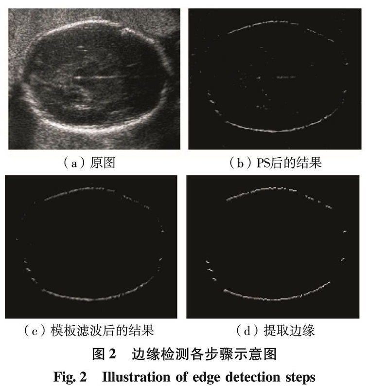
图2是边缘检测各步骤结果的示意图.其中,图2(a)是由RFs分类器得到的头围ROI; 图2(b)是对ROI求PS值的结果,PS值相对较大处对应着头骨的边缘部分; 图2(c)是对ROI模板滤波的结果,滤波模板是根据胎儿头围ROI中噪声的分布而设计的,如式(10),其中, e1(x,y)=(x-x0)2/a2+(y-y0)2/b2, e2(x,y)=(x-x0)2/(0.6a)2+(y-y0)2/(0.6b)2, 且(x0, y0)是全图的中心点, a和b分别是图像的长和宽的一半; 图2(d)是对图2(c)进行非极大值抑制,双阈值门限和形态学开运算后得到的头围边缘.
H(x,y)={0, e1(x,y)≥0.9, e2(x,y)≤1.2
1, e1(x,y)<0.9, e2(x,y)>1.2(10)
图2 边缘检测各步骤示意图
Fig.2 Illustration of edge detection steps